
Operational Principle
Butterfly wing as an imaging sensor
Butterflies are wonderful creatures with often shiny, colorful wings attracting attention of bystanders (Fig. 1). The secret of their coloration is frequently hidden in the intricate structures of tiny scales covering the whole surface of wings (Fig. 2). This is the layer of small particles that can be easily peeled and looks like dust on the fingertips. If magnified by the optical microscope, you can see that scales are positioned like shingles covering a roof of the house. They are small, plate-like structures, densely standing side by side, embedded in the wing by a petal few microns thick (Fig. 3). Scales are colored, but the reason for that can be seen only if scales are strongly magnified by an electron microscope (Fig. 2). Then we can observe intricate ridges and plates forming a complex optical structure – diffraction grating. Light, which is an electromagnetic wave, strongly and selectively interacts with grating so that some colors are reflected more strongly than others, and thus producing, so called, structural coloration. This is the kind of color that does not depend on the chemical composition of the wing, but rather on its internal architecture.
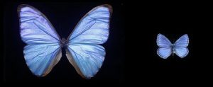
Fig. 1: Shiny blue Morpho (left) and polyommatus (right) butterflies
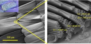
Fig. 2: Left – Scanning electron microscope image of scales covering the wing of Morpho didius butterfly (top-left inset shows optical image of a single scale). Right – nanostructures of individual wing scale.
Apart from strongly interacting with light, such structures are so small that only a few molecules of surrounding air can be packed inside ridges (Fig. 3). That is why, at this scale, butterfly wing scales interact with individual molecules, rather than with the gas as a whole. In such situation curious effects arise when light, surrounding gas and wing scale mutually interact. Wing scales absorb some amount of light, and slightly heat on the irradiated side. As a result, individual molecules pick-up more energy from the heated side than from the cooler one, and this difference creates a mechanical pressure acting on the scale. This force is small, but so is the mass of the scale, and the resulting effect is strong enough to be recorded by holography.
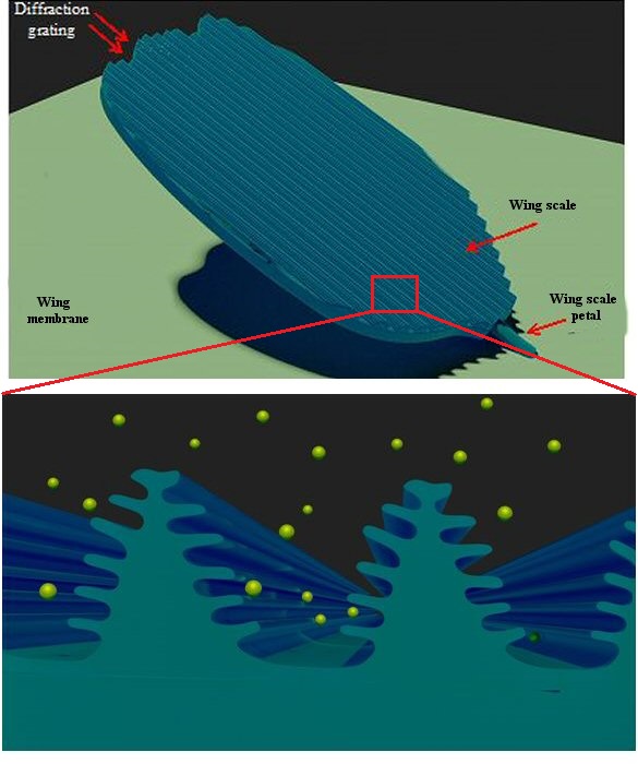
Fig. 3: A scheme of an individual butterfly wing scale is shown in the upper image. Cross-section of butterfly scale ridges (shown in blue color in the lower image), whose characteristic dimension is of the order of average distance of molecules (yellow spheres) at atmospheric pressure.
This kind of effect (now called photophoresis) was observed more than 150 years ago by the William Crookes, who designed a windmill-like structure and enveloped it in a transparent flask which was evacuated (Fig. 4). If irradiated with light, windmill starts turning with angular speed proportional to the light intensity. Initially, it was regarded as a proof of pressure of light, but with the gas molecules. In order to see the Crookes effect, the pressure has to be just right, so that the average distance between molecules of the surrounding gas is comparable to the thickness of the structure. In the case of the optical windmill this is of the order of 1/10 mm, and the gas pressure should be really low.
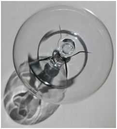
Fig. 4: Crookes radiometer: a windmill-like device placed in a vacuum flask. Upon irradiation windmill vanes rotate.
The main advantage of using wing-scale type of structure is that the effect can be observed without reducing the air pressure (vacuum), directly in the air. This is exactly what we did – we cut a circular piece of the Morpho butterfly wing and mounted it in an appropriate holder (Fig. 5). If observed more closely we can see that wing scales have a rather regular arrangement (rows and columns) and look just like pixels on a camera chip.
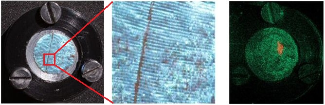
Fig.5: Image on the left shows a circular section of a Morpho butterfly wing, whose enlarged part is (middle) reveals an array of blue wing scales. Image on the right is a holographic reconstruction of a wing scale, where red patch is due to the irradiation with an infrared beam.
If a part of the wing is irradiated with a light beam, we can expect that each wing scale will slightly heat-up and move in proportion to the photophoretic forces. Holography is capable of revealing heated area and displaying it as an image (rightmost image in Fig. 5).
Our experiments have shown that butterfly wing can be used as a sensor in broad spectral range, spanning ultraviolet, visible, infrared and even terahertz radiation. Sensitivity and response speed are high, promising a new type of photo-detection for a range of applications where invisible radiation should be imaged.
Employing nanotechnology we will try to manufacture the pixels inspired by butterfly wing structures and to use those pixels in multispectral camera with holographic read-out.
(text and pictures: D. Pantelić)